Doctors at Ho Chi Minh City University Medical Center have successfully performed a complex cardiac procedure combining aortic valve replacement and coronary artery bypass using a full 3D endoscopic technique - the first case of its kind in Vietnam.
The hospital confirmed the operation on January 12, noting its significance as a milestone in minimally invasive cardiac surgery in the country.
The patient, a 64-year-old woman, was admitted with shortness of breath and chest pain during exertion. Her medical history included prior chest surgery and radiation therapy for breast cancer over 30 years ago.
Echocardiography revealed severe aortic regurgitation due to valve degeneration and 90% narrowing of the origin of the right coronary artery.
According to guidelines from the European Society of Cardiology and the American Heart Association, the recommended treatment was surgical aortic valve replacement combined with coronary artery bypass.
However, because of her past chest radiation, the traditional sternotomy approach posed heightened risks due to tissue adhesions and challenges in surgical field control.
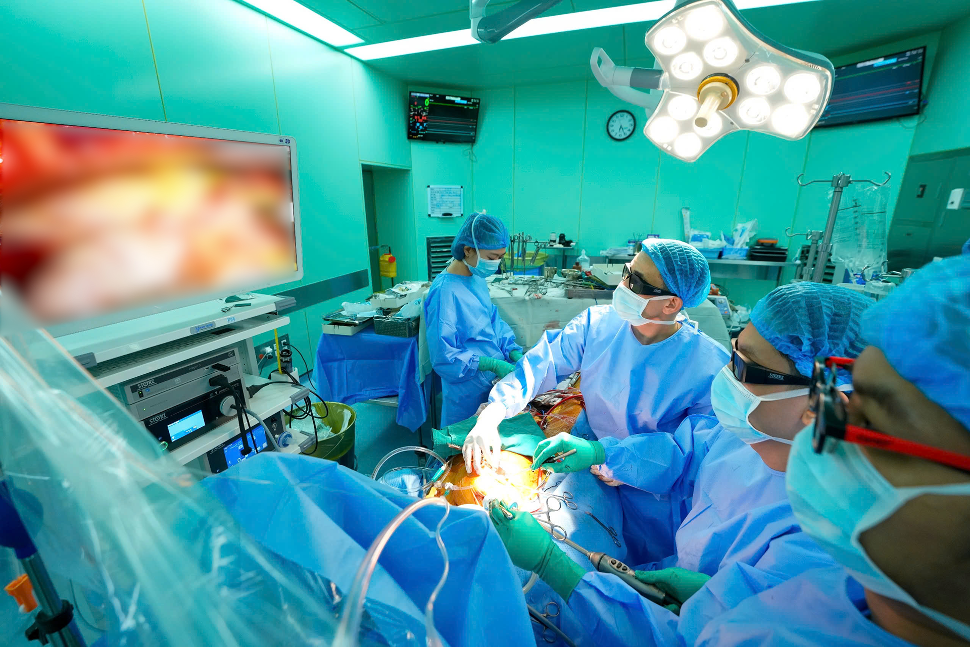
Following multidisciplinary consultation, the surgical team opted for a full 3D endoscopic approach, aiming to reduce trauma and support faster recovery.
High-tech surgery, seamless recovery
During the operation, the team simultaneously performed two tasks: implantation of a biological aortic valve and coronary artery bypass using a saphenous vein graft.
The cardiopulmonary bypass and cardiac arrest durations were well-controlled and comparable to those in traditional open-heart surgery.
Three hours after the procedure, the patient was extubated, and within 24 hours, echocardiography confirmed a 60% ejection fraction, proper valve function, and no regional wall motion abnormalities.
Dr. Pham Tran Viet Chuong, from the Department of Cardiovascular Surgery, explained that the 3D endoscopic system, equipped with a high-resolution camera, enables surgeons to visualize cardiac structures such as the aortic root, valves, and coronary arteries in fine detail - thanks to enhanced depth perception and image magnification.
“This technology significantly supports high-precision tasks like suturing a prosthetic valve or creating a coronary graft anastomosis within the confined space of endoscopic surgery,” Dr. Chuong said.
Pushing boundaries in cardiac care
Professor Dr. Nguyen Hoang Dinh, the hospital’s Deputy Director, emphasized that 3D endoscopic surgery offers an effective minimally invasive alternative for selected cardiac patients.
“When appropriately indicated and executed through close interdisciplinary collaboration, minimally invasive techniques can reduce complications and shorten recovery time,” he noted.
The success of this case opens new possibilities for advancing minimally invasive cardiac procedures in Vietnam, especially for high-risk patients previously deemed unsuitable for surgery due to complex medical histories.
Phuoc Sang